
Endoscopy vs Colonoscopy: Navigating the Differences in Gastrointestinal Imaging

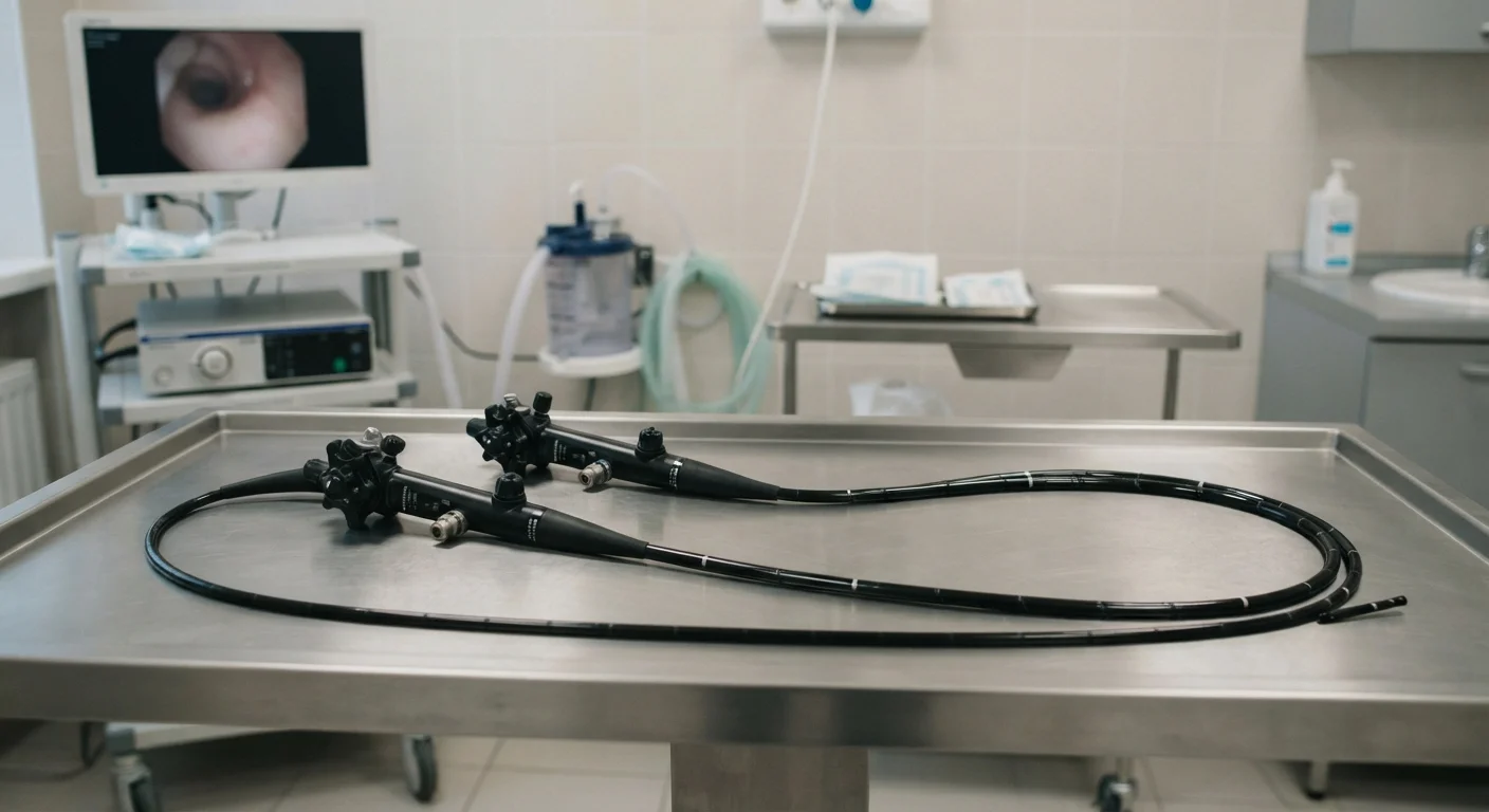
Photo: HerbHealWellness.com | Modern Guide To Wellness editorial
Key Takeaways
- Endoscopy examines the upper digestive tract; colonoscopy focuses on the large intestine and rectum.
- Both procedures use a thin, flexible tube with a camera called an endoscope, but the instruments differ in length and design.
- Colonoscopy requires a full bowel preparation; upper endoscopy requires only fasting.
- Both are performed under sedation and typically completed in under an hour.
- Clinicians may recommend one, both, or neither depending on a patient's specific symptoms and history.
What Each Procedure Actually Does
Both upper gastrointestinal endoscopy — often called an EGD (esophagogastroduodenoscopy) — and colonoscopy fall under the broad umbrella of endoscopic procedures. Each uses a thin, flexible tube equipped with a light and miniature camera to transmit real-time images to a monitor. The critical distinction lies in where that tube travels.
An upper endoscopy is inserted through the mouth and guided down the esophagus into the stomach and the first segment of the small intestine (the duodenum). A colonoscope, by contrast, is a longer, wider instrument introduced through the rectum and advanced through the full length of the large intestine, or colon.
Because each instrument accesses a different region, they answer different clinical questions. Clinicians use upper endoscopy to investigate symptoms originating in the upper digestive tract, while colonoscopy addresses lower GI concerns and serves as a primary colorectal cancer screening method. Just as imaging modalities target different anatomical structures — as explored in our comparison of MRI and CT scans — endoscopic tools are matched to specific diagnostic targets.
| Criterion | Upper Endoscopy (EGD) | Colonoscopy |
|---|---|---|
| Area examined | Esophagus, stomach, duodenum | Large intestine and rectum |
| Entry point | Through the mouth | Through the rectum |
| Instrument length | Approx. 100–125 cm | Approx. 130–160 cm |
| Preparation required | Fasting only (6–8 hours) | Full bowel cleanse plus fasting |
| Typical duration | 15–30 minutes | 30–60 minutes |
| Primary screening use | Upper GI symptoms, ulcers, celiac disease | Colorectal cancer, polyp detection |
| Biopsy capability | Yes | Yes |
| Polyp removal | Uncommon but possible | Standard and routine |
Preparation, Sedation, and the Patient Experience
Preparation requirements differ significantly between the two procedures, and this is often the most patient-relevant distinction.
For an upper endoscopy, patients are typically asked to fast for six to eight hours beforehand so the stomach is empty and the camera has a clear view. No bowel cleansing is needed.
Colonoscopy preparation is considerably more involved. Patients must follow a clear-liquid diet the day before and consume a prescribed laxative solution — sometimes called a bowel prep — to fully cleanse the colon. Many patients report that this preparation is the most uncomfortable part of the entire process.
Both procedures are almost always performed under conscious sedation or monitored anesthesia, meaning patients are relaxed and minimally aware during the examination. The procedures themselves are generally brief — upper endoscopy typically takes 15 to 30 minutes, while a colonoscopy may take 30 to 60 minutes depending on findings.
19M+
Colonoscopies performed annually in the US
The American Society for Gastrointestinal Endoscopy estimates more than 19 million colonoscopies are performed each year in the United States.
~7M
Upper endoscopies performed annually in the US
Upper GI endoscopy is among the most frequently performed GI procedures in the US, according to gastroenterological utilization data.
<1%
Major complication rate for both procedures
Serious complications such as perforation or major bleeding occur in fewer than 1 in 1,000 procedures for both EGD and colonoscopy in standard clinical settings.
Recovery involves a short observation period to allow sedation to wear off, and patients require a companion to drive them home. Minor bloating or gas is common afterward; both resolve quickly.
Risks, Capabilities, and When Both May Be Needed
Both procedures carry a small risk of complications, including bleeding, infection, or — very rarely — perforation of the digestive tract wall. These risks are low in the hands of a trained gastroenterologist, but clinicians weigh them against the diagnostic benefit for each individual patient.
A major advantage of endoscopic examination is its therapeutic capability. Unlike passive imaging, both EGD and colonoscopy allow the physician to take biopsies, remove polyps, control minor bleeding, or dilate narrowed segments during the same session.
Capsule Endoscopy: A Non-invasive Alternative
In some cases, a patient may need both procedures — for example, when symptoms span both the upper and lower GI tract, or when a complete evaluation is required before a planned surgery. Occasionally both are performed during a single anesthesia session for efficiency.
The decision about which procedure — or whether either is appropriate — belongs to a qualified gastroenterologist or primary care physician who can assess the full clinical picture. Readers experiencing persistent digestive symptoms should consult a healthcare professional rather than attempting to self-select a procedure.
This article is for general informational purposes only and does not constitute medical advice. Always consult a qualified healthcare provider for guidance on your individual health circumstances.
The content on this site is for informational purposes only and is not a substitute for professional advice. Always consult a qualified professional for guidance specific to your situation.