
MRI vs CT Scan: What Each One Reveals and When Clinicians Choose Between Them

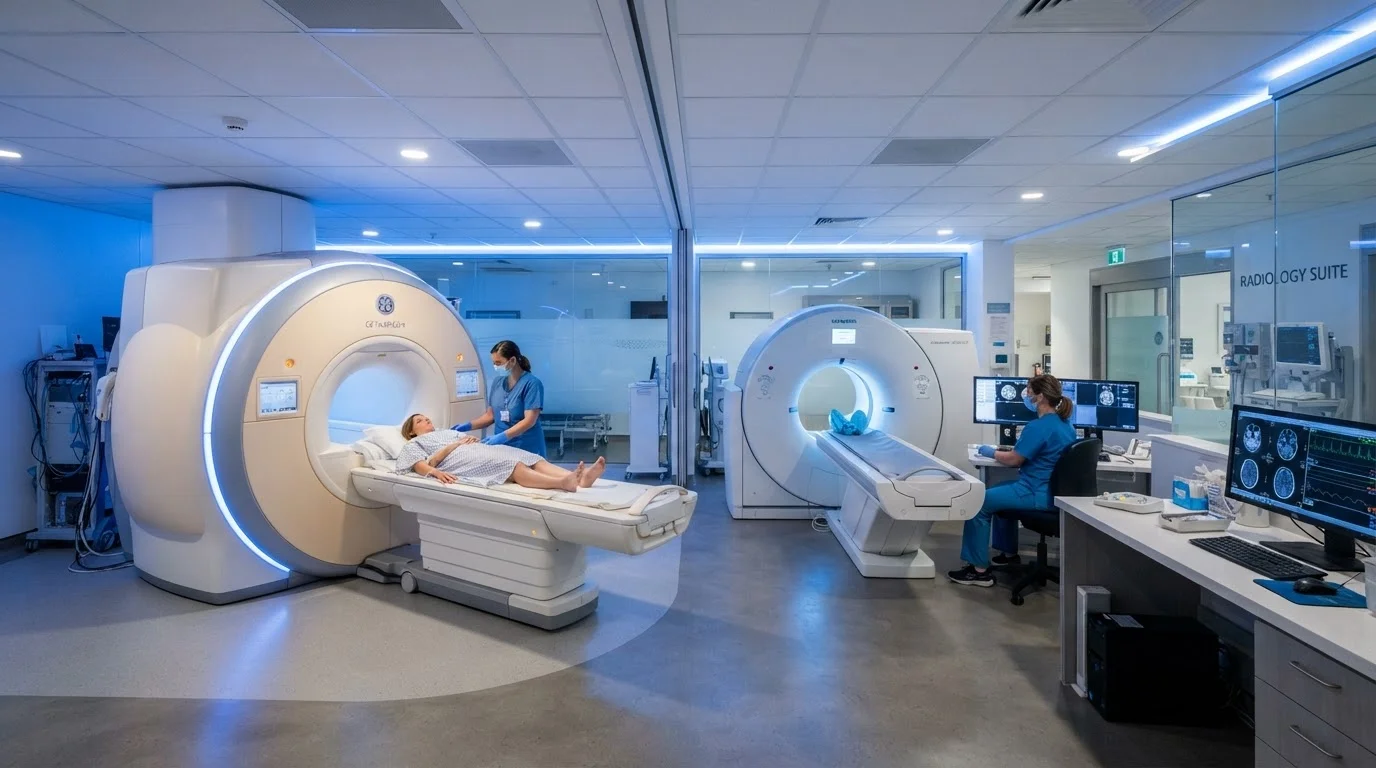
Photo: HerbHealWellness.com | Modern Guide To Wellness editorial
Key Takeaways
- MRI uses magnetic fields and radio waves; CT uses ionizing X-ray radiation to generate images.
- CT is faster and better suited to emergencies, bone injuries, and lung assessment.
- MRI provides superior soft-tissue contrast for neurological, musculoskeletal, and pelvic conditions.
- CT exposes patients to ionizing radiation; MRI does not, though MRI has contraindications involving metal implants.
- Clinicians choose between them based on anatomy, urgency, patient safety factors, and clinical question.
How Each Technology Works
Despite both producing cross-sectional images of the body's interior, MRI and CT operate on entirely different physical principles.
MRI uses a powerful magnetic field — typically 1.5 to 3 Tesla in clinical settings — along with radiofrequency pulses to cause hydrogen atoms in body tissue to emit detectable signals. Because different tissues (fat, water, muscle, nerve) contain hydrogen in varying concentrations, the scanner can produce images with exceptional soft-tissue contrast. Crucially, MRI involves no ionizing radiation.
CT scanning rotates an X-ray source around the patient while detectors capture the attenuated beams from multiple angles. A computer reconstructs these measurements into detailed cross-sectional slices. CT is considerably faster than MRI — a full chest-abdomen-pelvis scan may take under a minute on modern equipment — but it does involve ionizing radiation. For perspective on what that means clinically, see our article on radiation exposure in medical imaging.
| Criterion | MRI | CT Scan |
|---|---|---|
| Underlying technology | Magnetic field and radio waves | Ionizing X-ray radiation |
| Ionizing radiation | None | Yes (low to moderate dose) |
| Scan duration | 20–60 minutes typically | Seconds to a few minutes |
| Soft-tissue contrast | Excellent | Moderate |
| Bone and calcification detail | Limited | Excellent |
| Best clinical uses | Brain, spine, joints, pelvis | Trauma, lungs, abdomen, bones |
| Metal implant contraindication | Yes — many implants excluded | Generally no restriction |
| Typical emergency availability | Limited; longer setup time | Widely available; rapid access |
What Each Scan Reveals Best
The physical principles underlying each modality translate directly into clinical strengths and limitations.
MRI excels at:
- Brain and spinal cord pathology — including tumors, stroke, and demyelinating conditions
- Soft-tissue structures such as ligaments, cartilage, and intervertebral discs
- Pelvic organs, including the uterus, ovaries, and prostate
- Detecting subtle lesions where contrast between tissue types matters most
CT excels at:
- Bone structure and fracture assessment
- Pulmonary conditions — lung nodules, pneumonia, pulmonary embolism
- Abdominal emergencies including appendicitis, bowel obstruction, and organ lacerations
- Rapid triage in polytrauma situations
~40M
MRI procedures performed annually in the US
The FDA estimates approximately 40 million MRI examinations are performed each year in the United States, reflecting its central role in diagnostic imaging.
~85M
CT scans performed annually in the US
The National Cancer Institute cites roughly 85 million CT scans per year in the US, making CT the most frequently used advanced imaging modality.
It is worth noting that imaging findings always require clinical interpretation. As our editorial team explores separately, imaging alone cannot diagnose most conditions — scans provide structural information that clinicians combine with history, examination, and laboratory results.
Safety Considerations and Contraindications
Both modalities are generally safe, but each carries distinct considerations that influence which is appropriate for a given patient.
MRI contraindications center on the strong magnetic field. Patients with certain cardiac pacemakers, cochlear implants, ferromagnetic aneurysm clips, or other metallic implants may be ineligible — or require device-specific clearance before scanning. MRI is also considerably louder and more time-consuming, which can affect patients with claustrophobia. Gadolinium-based contrast agents used in some MRI protocols carry a small risk of adverse reactions and require caution in patients with impaired kidney function.
CT radiation is the primary safety consideration for that modality. While a single CT scan delivers a dose that is considered low in absolute terms, cumulative exposure from repeated imaging is a factor clinicians weigh — particularly for pediatric patients or individuals requiring frequent monitoring. Iodinated contrast used in CT also carries allergy and renal risks comparable to gadolinium.
Contrast Agents: An Important Distinction
Patients who are pregnant, very young, or managing chronic conditions should discuss the specific risks and benefits with their clinical team before undergoing either type of scan. Always consult a qualified healthcare professional for guidance specific to your circumstances.
How Clinicians Decide Which to Order
The choice between MRI and CT is rarely arbitrary. Clinicians weigh several intersecting factors:
- The clinical question: What tissue or structure needs to be evaluated, and at what level of detail?
- Urgency: CT's speed makes it the default in acute trauma or hemodynamic instability where minutes matter.
- Patient-specific factors: Metal implants, renal function, pregnancy, and claustrophobia all constrain options.
- Availability: MRI scanners require longer booking windows at many facilities; CT is more rapidly accessible in most emergency departments.
- Radiation profile: Minimizing cumulative dose may favor MRI when either modality would answer the clinical question equally well.
Before your scan, it is worth knowing what to expect and what questions to raise with your care team. Our guide on preparing for a diagnostic procedure covers exactly that ground.
This article is for general informational and educational purposes only and does not constitute medical advice. Always consult a qualified healthcare professional regarding any medical imaging or health decisions specific to your situation.
The content on this site is for informational purposes only and is not a substitute for professional advice. Always consult a qualified professional for guidance specific to your situation.