
Radiation Exposure in Medical Imaging: Separating Real Risk from Common Fear

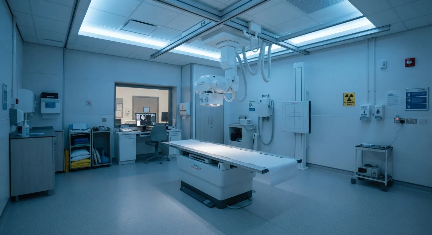
Photo: HerbHealWellness.com | Modern Guide To Wellness editorial
Key Takeaways
- Radiation doses from most routine imaging are far lower than many patients assume.
- Background radiation from the natural environment exposes everyone to ionizing radiation daily.
- The clinical benefit of an ordered scan almost always outweighs the small statistical risk.
- CT scans deliver higher doses than X-rays, but dose varies significantly by body region and protocol.
- Declining a clinically necessary scan carries its own health risks that should be weighed carefully.
Understanding Dose: Context Is Everything
When a clinician orders a chest X-ray or CT scan, many patients feel a spike of anxiety about radiation. That concern is understandable — ionizing radiation is associated with cancer risk, and the association is real. What is frequently misunderstood, however, is scale. Radiation dose is measured in millisieverts (mSv), and the doses delivered by most diagnostic imaging fall far below the thresholds associated with measurable harm.
Every person in the United States is exposed to background radiation continuously — from radon in soil, cosmic rays at altitude, and naturally occurring radioactive materials in food and building materials. Understanding that medical imaging doses are typically comparable to days or weeks of this unavoidable background exposure reframes the conversation significantly.
~3 mSv
Average annual background radiation dose in the U.S.
According to the National Council on Radiation Protection and Measurements, natural background sources account for the majority of average American radiation exposure.
0.1 mSv
Effective dose from a standard chest X-ray
The American College of Radiology estimates this is roughly equivalent to 10 days of natural environmental radiation exposure for an average U.S. resident.
~50%
Share of U.S. radiation exposure from medical imaging
The National Council on Radiation Protection and Measurements has reported that medical imaging accounts for roughly half of total radiation exposure for the average American.
If you are preparing for an upcoming scan and want to know the right questions to ask beforehand, see Preparing for a Diagnostic Procedure: Questions Worth Asking Before You Go.
Common Myths About Imaging Radiation — Corrected
Misconceptions about medical radiation are widespread and, in some cases, lead patients to refuse or delay imaging that could significantly affect their care. The following myth-and-fact pairs address the most common misunderstandings, grounded in guidance from radiology and health physics organizations.
Myth
Any radiation exposure from medical imaging is dangerous and should be avoided.
Fact
Low-level ionizing radiation from diagnostic imaging carries a very small statistical risk, and the clinical benefit of accurate diagnosis almost always outweighs that risk.
Ionizing radiation can damage DNA, which is why people associate it with cancer risk. However, dose matters enormously. A standard chest X-ray delivers roughly 0.1 millisieverts (mSv) of effective dose — equivalent to about 10 days of natural background radiation exposure. The U.S. average annual background dose is approximately 3 mSv from sources such as radon gas, cosmic rays, and soil. At diagnostic imaging doses, the increased cancer risk is either immeasurably small or theoretical, estimated through extrapolations from much higher-exposure events rather than direct evidence from imaging populations.
Myth
A CT scan is essentially the same as an X-ray in terms of radiation.
Fact
CT scans deliver significantly higher radiation doses than plain X-rays — often 10 to 100 times more depending on the region imaged — which is why they are ordered selectively.
A conventional chest X-ray uses a single pass of a focused beam, whereas a computed tomography (CT) scan acquires hundreds of cross-sectional images using a rotating X-ray source. An abdominal and pelvic CT, for example, may deliver an effective dose in the range of 8–14 mSv, compared to 0.02 mSv for a hand X-ray. Radiologists and clinicians apply the ALARA principle — As Low As Reasonably Achievable — adjusting scan parameters to minimize dose while preserving diagnostic quality. To understand when a CT is chosen over an MRI, see MRI vs CT Scan: What Each One Reveals.
Myth
Radiation exposure accumulates permanently in the body from scans.
Fact
Ionizing radiation from imaging passes through the body; it does not remain stored or "build up" in tissues over time.
Unlike some contrast agents or radioactive tracers used in nuclear medicine, the X-ray photons used in conventional imaging and CT scans pass through the body and are detected by sensors on the other side. They do not deposit a substance inside you. What accumulates statistically over a lifetime of scans is biological risk — the small probability that radiation-induced cellular changes could contribute to cancer years later. This risk remains low at diagnostic dose levels, but it is the reason clinicians track a patient's imaging history and avoid redundant scans.
Myth
Patients should request scans proactively to catch problems early.
Fact
Scans ordered without a clinical indication can produce false positives, lead to unnecessary procedures, and add avoidable radiation burden — they are not routine wellness tools.
Whole-body CT scans marketed directly to consumers are not endorsed by major radiology or preventive medicine organizations for asymptomatic individuals, precisely because incidental findings are common and often require invasive follow-up that carries its own risks. Imaging is most valuable as part of a clinical reasoning process — not as a standalone screening tool for most conditions. For context on how imaging fits into a broader diagnostic picture, see Why Imaging Alone Cannot Diagnose Most Conditions.
Myth
Lead aprons completely protect all sensitive organs during any scan.
Fact
Lead aprons shield areas outside the imaging field but offer little protection against internal scatter, and their use depends on the region being imaged and current clinical protocols.
Lead shielding can reduce exposure to radiosensitive organs — such as the thyroid or reproductive organs — when they fall near the edges of a scan field. However, modern protocols and guidelines from organizations such as the American College of Radiology have evolved. In some contexts, poorly placed shielding can obscure diagnostic information or cause automatic exposure compensation that actually increases dose. Facilities calibrate shielding practices to balance protection with image quality, so practices vary and are guided by updated evidence rather than a universal rule.
Never Decline a Necessary Scan Without Consulting Your Clinician
This article is for informational purposes only and is not a substitute for professional medical advice. Always consult a qualified healthcare provider with questions about imaging recommendations or radiation safety specific to your situation.
Weighing Risk Against Clinical Benefit
Radiation risk from diagnostic imaging is real but very small — and it must always be weighed against the clinical benefit of obtaining the information a scan provides. A clinician who orders an abdominal CT for suspected appendicitis is weighing a small statistical radiation risk against the much more immediate risk of a missed diagnosis. In that context, the scan is almost always the safer option.
Special Considerations for Pregnancy
Clinicians and medical physicists apply the ALARA principle to keep imaging doses as low as diagnostic quality will allow. Facilities regularly audit protocols, calibrate equipment, and adopt newer technologies — such as iterative reconstruction algorithms in CT — that reduce dose without sacrificing image clarity. For a broader perspective on how diagnostic tools combine with clinical judgment, see Why Imaging Alone Cannot Diagnose Most Conditions.
Being an informed patient means understanding that declining a scan is itself a clinical decision with consequences — not a neutral, risk-free choice. Engage your care team in open conversation about why imaging has been recommended, what alternatives exist, and what information the scan is expected to provide.
The content on this site is for informational purposes only and is not a substitute for professional advice. Always consult a qualified professional for guidance specific to your situation.