
Why Imaging Alone Cannot Diagnose Most Conditions

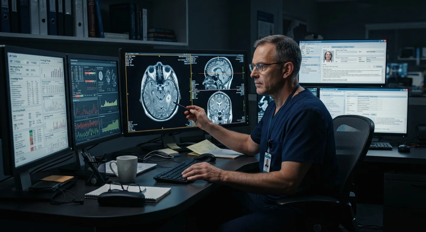
Photo: HerbHealWellness.com | Modern Guide To Wellness editorial
Key Takeaways
- Imaging reveals anatomy and structure, but cannot independently establish a clinical diagnosis in most cases.
- Clinicians integrate scan findings with patient history, physical exams, and laboratory results to reach conclusions.
- A normal scan does not always mean nothing is wrong, and an abnormal scan does not always mean something is.
- Radiologists report what they observe; treating clinicians interpret that report within the full clinical picture.
- Many conditions — including functional disorders — produce no visible changes on any imaging modality.
What Imaging Actually Shows — and What It Doesn't
Medical imaging technology — X-rays, CT scans, MRI, and ultrasound — has transformed clinical medicine by allowing clinicians to see inside the body without surgery. Yet these tools have a fundamental limitation that patients often underestimate: they capture structure, not function, causation, or clinical significance on their own.
A scan can reveal that a mass is present, that a bone is fractured, or that fluid has accumulated around the heart. What it cannot do is tell the clinician why these findings exist, whether they are causing the patient's symptoms, or what treatment is appropriate — without additional context. To learn how different modalities compare in what they reveal, see our overview of MRI vs CT scanning.
This gap between observation and interpretation is why diagnosis is always a synthesis — not a single data point.
Myth
If my scan is clear, there's nothing wrong with me.
Fact
A normal scan rules out structural abnormalities visible to that modality — it does not rule out illness entirely.
Many conditions are functionally rather than structurally based. Migraine, functional gastrointestinal disorders, early-stage autoimmune conditions, and certain neuropathies may produce significant symptoms without any detectable imaging abnormality. A negative scan narrows the differential diagnosis but does not end the clinical investigation.
Myth
A scan gives the doctor a definitive diagnosis instantly.
Fact
Imaging provides data that must be interpreted alongside the patient's clinical presentation, history, and other test results.
Radiology reports use language like "consistent with," "suggestive of," or "cannot exclude" deliberately — because the image alone rarely establishes certainty. A shadow on a chest X-ray could represent pneumonia, a tumor, scarring, or an artifact. Context — symptoms, exposure history, lab markers — determines which interpretation is most defensible. The evolution of diagnostic tools reflects decades of learning that no single instrument is sufficient alone.
Myth
More imaging always leads to better, faster diagnoses.
Fact
Unnecessary imaging can introduce incidental findings, radiation exposure, and diagnostic delay without improving accuracy.
Ordering imaging without a clear clinical question in mind can generate more uncertainty than clarity. Incidental findings — structures that look unusual but are clinically irrelevant — require follow-up investigations, create patient anxiety, and consume healthcare resources. Clinical guidelines from bodies such as the American College of Radiology help clinicians determine when imaging adds genuine diagnostic value. For context on radiation considerations, see our article on radiation exposure in medical imaging.
Myth
Radiologists are the ones who diagnose conditions from scans.
Fact
Radiologists interpret and report imaging findings; diagnosis is the responsibility of the treating clinician synthesizing all available data.
Radiology is a consultation specialty. A radiologist provides a structured report describing observed findings and, where appropriate, a differential — a ranked list of possible explanations. The treating physician or specialist then integrates that report with the clinical picture. In some settings, such as emergency medicine, clinicians may review images directly, but radiologist review remains a separate and distinct step. Diagnosis is ultimately a clinical act, not a purely technological one.
Myth
Ultrasound and MRI work the same way — they're just different brands of scanner.
Fact
Each imaging modality uses a different physical mechanism and is suited to different clinical questions.
Ultrasound uses high-frequency sound waves reflected off tissue boundaries — it excels at visualizing soft-tissue structures and fluid in real time, with no ionizing radiation. MRI uses magnetic fields and radio waves to generate detailed soft-tissue contrast. CT uses X-rays processed computationally to produce cross-sectional images suited to trauma and vascular structures. Each modality has distinct strengths, limitations, and clinical indications. See our guide to ultrasound in clinical practice for a deeper look at one commonly misunderstood tool.
How Clinicians Build a Complete Diagnostic Picture
In clinical practice, imaging results are one layer within a structured diagnostic framework. That framework typically includes:
- Patient history: Duration, severity, and pattern of symptoms; relevant past diagnoses; medications; family history.
- Physical examination: Direct assessment of the body that may reveal findings invisible to imaging — tenderness, reflexes, organ enlargement, or neurological signs.
- Laboratory data: Blood counts, inflammatory markers, metabolic panels, and pathology results that contextualize what a scan shows.
- Clinical probability: Experienced clinicians apply probabilistic reasoning — given this patient's age, risk factors, and presentation, how likely is each possible explanation?
A radiologist's report describes what the imaging shows but does not carry the full burden of diagnosis. The treating clinician must weigh that report against everything else known about the patient. This is especially relevant for chronic conditions, where imaging findings may predate symptoms by years or persist after symptoms resolve.
Don't Rely Solely on Imaging Results
Why This Matters for Patients
Understanding imaging's role — and its limits — helps patients engage more productively with their care. If a scan comes back normal, that result is genuinely useful information, but it does not close the diagnostic conversation. Many conditions, including irritable bowel syndrome, fibromyalgia, and certain cardiac arrhythmias, produce no visible structural change on any imaging modality.
Conversely, imaging frequently detects incidental findings — abnormalities unrelated to a patient's symptoms that require careful clinical judgment to interpret. Not every finding on a scan demands intervention. The diagnostic equipment glossary covers terms like "incidentaloma" that patients may encounter in radiology reports.
Clinicians order imaging strategically, not as a comprehensive sweep. Understanding why a specific modality was chosen — and what question it is intended to answer — is a reasonable and productive thing to ask your care team.
~30%
Incidental findings rate in abdominal CT scans
Research published in peer-reviewed radiology literature has estimated that incidental findings appear in roughly 30% of abdominal CT scans performed for unrelated indications, underscoring the complexity of scan interpretation.
4–6 data sources
Inputs clinicians typically integrate per diagnosis
Clinical decision-making frameworks in academic medicine routinely describe combining history, examination, labs, imaging, and risk stratification tools before reaching a working diagnosis.
This article is for general informational and educational purposes only and does not constitute medical advice. Always consult a qualified healthcare professional for guidance about your specific health situation.
The content on this site is for informational purposes only and is not a substitute for professional advice. Always consult a qualified professional for guidance specific to your situation.