
The Electrocardiogram Explained: Reading the Story Your Heart Tells

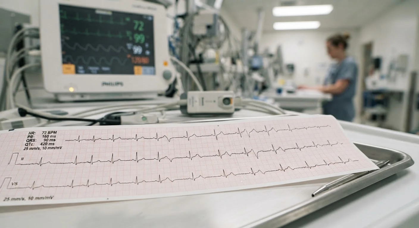
Photo: HerbHealWellness.com | Modern Guide To Wellness editorial
Key Takeaways
- An ECG records the electrical activity of the heart through electrodes placed on the skin — no needles or radiation involved.
- The characteristic waves (P, QRS, T) each correspond to a specific phase of the heart's electrical cycle.
- ECGs can detect arrhythmias, signs of a heart attack, conduction problems, and structural abnormalities.
- A standard 12-lead ECG takes only a few minutes and is a cornerstone of cardiac diagnosis.
- Abnormal findings always require interpretation by a qualified clinician in context with the patient's full history.
What an ECG Actually Measures
Every heartbeat begins with an electrical impulse that originates in the heart's natural pacemaker — the sinoatrial (SA) node — and travels through a precisely ordered conduction pathway. This electrical activity causes the heart muscle to contract in a coordinated sequence, pumping blood through the chambers and out to the body.
An ECG captures this electrical journey from the surface of the skin. Electrodes — small adhesive patches — are placed at standardized positions on the chest, arms, and legs. Each electrode detects the tiny voltage changes generated as the electrical wavefront passes through different regions of the heart muscle. The machine amplifies these signals and plots them as a continuous wave over time.
Importantly, an ECG measures electrical activity, not mechanical pumping directly. A separate test, such as an echocardiogram, is needed to visualize how the heart moves. That said, electrical patterns reliably reflect much of what is happening structurally and functionally. See our guide to diagnostic lab equipment for how ECGs fit alongside other clinical tools.
~300 million
ECGs performed globally each year
The ECG is one of the most frequently performed cardiac tests worldwide, reflecting its central role in clinical evaluation across specialties.
10 seconds
Duration of the actual ECG recording
A standard 12-lead ECG recording captures only about ten seconds of electrical data — sufficient to assess rhythm, rate, and conduction in most clinical scenarios.
100+ years
ECG in continuous clinical use
The electrocardiogram has been a cornerstone of cardiac diagnosis since the early twentieth century, remaining highly relevant despite advances in imaging and molecular diagnostics.
Decoding the Waves: P, QRS, and T
The ECG tracing is not random noise — each peak and valley maps directly onto a phase of the heart's electrical cycle. Understanding the basic components helps demystify what clinicians are looking for.
- P wave: A small rounded bump representing atrial depolarization — the electrical activation of the upper chambers (atria) that causes them to contract and push blood into the ventricles.
- PR interval: The flat segment between the P wave and the next complex, reflecting the brief delay at the atrioventricular (AV) node that allows the ventricles to fill before they contract.
- QRS complex: The tall, sharp spike that dominates the tracing. It represents ventricular depolarization — the electrical activation of the lower chambers (ventricles) as they contract forcefully to pump blood to the lungs and body. Because the ventricles are much larger than the atria, this complex is far more prominent.
- ST segment: The plateau between the QRS complex and the T wave. Elevation or depression of this segment is a key marker clinicians look for when suspecting a heart attack.
- T wave: A broader wave reflecting ventricular repolarization — the electrical reset of the ventricles in preparation for the next beat.
By measuring the height, width, shape, and timing of these features, a clinician can infer a remarkable amount about how well the heart's electrical system is functioning.
“The ECG remains one of the most information-dense tests in medicine per unit of time and cost. No other ten-second test tells you as much about the heart's electrical architecture.”
— A clinical cardiologist and medical educator, Cardiologist and academic faculty member at a US medical school
The 12-Lead ECG: Viewing the Heart From Multiple Angles
A standard clinical ECG uses 12 leads — not 12 separate electrodes, but 12 different electrical viewpoints derived from the electrode placements. Each lead looks at the heart from a different angle, much like photographing a building from multiple positions to understand its full shape.
Six limb leads examine the heart in a vertical plane, while six precordial (chest) leads examine it horizontally. Together, they allow clinicians to pinpoint where in the heart an abnormality is occurring — for example, whether ST-segment changes appear in leads that correspond to the front wall of the heart versus the back or side walls. This localization is particularly critical when evaluating possible heart attacks, since different coronary arteries supply different territories.
Bring Previous ECGs to Appointments
For readers curious about the broader landscape of monitoring tools — including devices used at home — understanding key vital signs provides helpful context for how heart rate and rhythm fit within the full picture of cardiovascular health.
Common Reasons an ECG Is Ordered
Clinicians order ECGs across a wide range of clinical situations. While this is not an exhaustive list, the most common indications include:
- Chest pain or pressure — to assess whether symptoms may be cardiac in origin, including ruling in or out a myocardial infarction (heart attack).
- Palpitations or irregular pulse — to identify arrhythmias such as atrial fibrillation, supraventricular tachycardia, or heart block.
- Shortness of breath or syncope (fainting) — to check whether a cardiac rhythm or conduction problem is contributing.
- Routine preoperative screening — to establish a baseline and identify any underlying cardiac issues before surgery.
- Medication monitoring — certain drugs affect the QT interval (the time for ventricles to reset), requiring periodic ECG checks.
- Follow-up after a known heart condition — to monitor progression or treatment response.
It is worth noting that a normal ECG does not rule out all heart disease, and an abnormal ECG does not always indicate serious pathology. Clinical interpretation always depends on the individual patient's presentation and medical history.
Wearable ECGs: Useful, But Limited
This article is for informational purposes only and does not constitute medical advice. Always consult a qualified healthcare provider regarding any cardiac symptoms, test results, or treatment decisions.
Frequently Asked Questions
The content on this site is for informational purposes only and is not a substitute for professional advice. Always consult a qualified professional for guidance specific to your situation.