
Keratoconus: When the Cornea Changes Shape

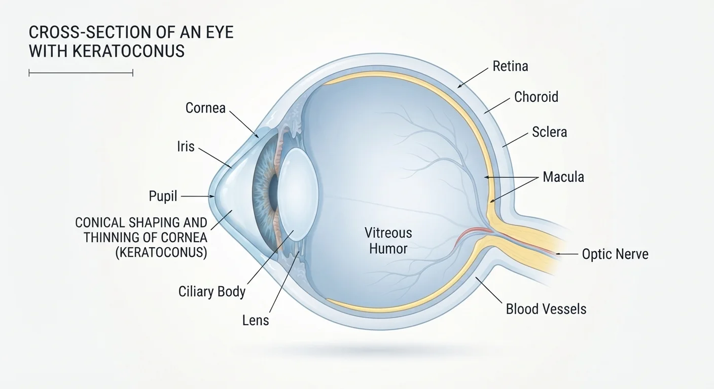
Photo: HerbHealWellness.com | Modern Guide To Wellness editorial
Key Takeaways
- Keratoconus causes the cornea to thin and bulge, distorting vision in ways glasses often cannot fully correct.
- Symptoms typically begin in the teens or early twenties and may progress over many years.
- Rigid or specialty contact lenses are often the most effective non-surgical approach for managing vision.
- Corneal collagen cross-linking is an established procedure that can slow or halt progression.
- Regular monitoring by an eye care professional is essential once the condition is diagnosed.
- Early diagnosis gives patients the most treatment options and the best chance of preserving vision.
What Happens to the Cornea
The cornea normally forms a smooth, symmetrical dome that bends incoming light precisely onto the retina. In keratoconus, collagen fibers within the corneal tissue weaken, causing the structure to thin unevenly and bow forward. The resulting cone shape scatters light rather than focusing it cleanly, producing the blurred and distorted images characteristic of the condition.
Unlike straightforward refractive errors such as myopia or astigmatism, keratoconus creates irregular astigmatism — the corneal surface warps in an unpredictable, asymmetric pattern that conventional corrective lenses struggle to compensate for. This is why many patients notice that new glasses prescriptions stop working well relatively quickly during active progression.
Recognizing the Symptoms
Keratoconus often announces itself subtly. Early signs include mildly blurred vision, increased sensitivity to light and glare, and a tendency to squint or tilt the head to find a clearer line of sight. Many people initially receive prescriptions for glasses or soft contact lenses before a definitive diagnosis is made.
As the condition advances, additional symptoms can emerge:
- Distortion or streaking around lights, particularly at night
- Multiple ghost images or "halos" around light sources
- Rapid prescription changes that provide only temporary relief
- One eye often affected more severely than the other
In more advanced stages, some individuals develop acute corneal hydrops — a sudden swelling caused by a break in the cornea's innermost layer — which causes rapid, painful vision loss. This is relatively uncommon but requires prompt evaluation.
When to Seek Prompt Evaluation
Who Is at Risk
Keratoconus affects an estimated 1 in 2,000 people in the general population, though some research suggests it may be more prevalent than previously recognized as diagnostic tools improve. It tends to emerge in the teens and early twenties and affects males and females across all ethnic backgrounds.
~1 in 2,000
Estimated prevalence in the general population
Commonly cited epidemiological estimates suggest keratoconus affects approximately 1 in 2,000 individuals, though newer diagnostic technologies may reveal higher rates.
Teens–20s
Typical age of onset
Most cases of keratoconus first present during adolescence or early adulthood, with progression often continuing into the thirties before stabilizing.
Several factors are associated with higher risk:
- Family history: First-degree relatives of those with keratoconus have a meaningfully elevated risk.
- Chronic eye rubbing: Repeated mechanical trauma may accelerate corneal thinning.
- Certain systemic conditions: Down syndrome, connective tissue disorders such as Marfan syndrome, and some allergic conditions are associated with higher prevalence.
- Poorly fitted contact lenses: Prolonged wear of ill-fitting lenses may contribute in susceptible individuals.
How Keratoconus Is Diagnosed
A routine eye examination may raise suspicion, but confirming keratoconus typically requires corneal topography or tomography — detailed mapping technologies that measure the curvature and thickness of the cornea across its entire surface. These maps reveal the irregular steepening pattern that distinguishes keratoconus from ordinary astigmatism.
In early cases, changes may be very subtle, which is why a comprehensive examination by an optometrist or ophthalmologist — rather than a standard vision screening — is important for anyone experiencing unexplained progressive visual distortion or frequent prescription changes.
Managing Keratoconus: From Lenses to Procedures
Treatment is guided by how far the condition has progressed and how well vision can be corrected. The range of vision correction options available includes several strategies specifically suited to keratoconus:
- Rigid gas-permeable (RGP) contact lenses: These hard lenses vault over the irregular corneal surface, creating a smooth optical surface. They are a cornerstone of keratoconus management for moderate to advanced cases.
- Scleral lenses: Larger-diameter rigid lenses that rest on the white of the eye (sclera) rather than the cornea itself, offering superior comfort and stability for highly irregular corneas.
- Hybrid lenses: A rigid center with a soft skirt, combining optical clarity with improved comfort.
- Corneal collagen cross-linking (CXL): An established procedure using riboflavin drops and ultraviolet light to strengthen corneal collagen bonds and slow or halt progression. It is not intended to reverse existing changes but to stabilize the cornea.
- Corneal transplant (keratoplasty): Reserved for advanced cases where contact lenses can no longer provide functional vision or significant scarring is present. Modern techniques — particularly deep anterior lamellar keratoplasty (DALK) — replace only the diseased corneal layers rather than the full thickness.
Ask About Specialty Lens Fitters
It is worth noting that keratoconus is quite different from age-related vision changes such as presbyopia, which involves the lens rather than the cornea and follows a predictable pattern with aging.
This article is for general informational purposes only and does not constitute medical advice. If you are experiencing vision changes or symptoms described here, consult a qualified eye care professional for a proper evaluation.
Frequently Asked Questions
The content on this site is for informational purposes only and is not a substitute for professional advice. Always consult a qualified professional for guidance specific to your situation.