
Diagnostic Equipment Through the Decades: How Clinical Tools Have Evolved

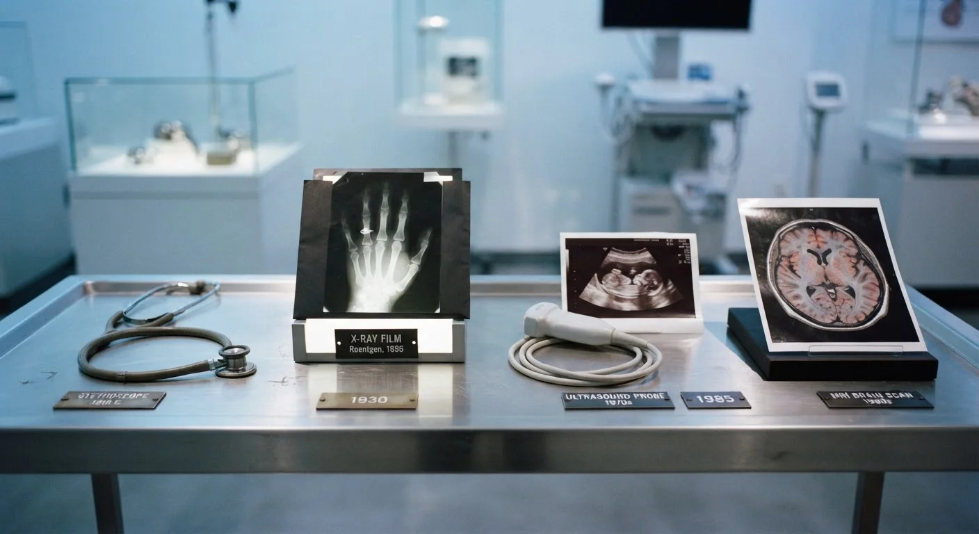
Photo: HerbHealWellness.com | Modern Guide To Wellness editorial
Key Takeaways
- Diagnostic tools have evolved from simple mechanical instruments to complex digital and AI-assisted systems over the past 200 years.
- Each technological leap — X-rays, ultrasound, MRI — expanded what clinicians could detect without invasive procedures.
- Modern diagnostics increasingly combine multiple data streams, including imaging, biomarkers, and wearable sensor data.
- Understanding how these tools work helps patients engage more confidently with their own clinical care.
- No single device or test provides a complete picture; clinicians always interpret results in broader clinical context.
Two Centuries of Clinical Insight
The instruments clinicians use to peer inside the human body have undergone a series of profound transformations — each driven by advances in physics, chemistry, computing, and materials science. What began with a rolled-up paper tube has expanded into a universe of digital sensors, cross-sectional imagers, and machine-learning algorithms trained on millions of scans.
Understanding this progression matters beyond historical curiosity. When patients encounter unfamiliar machines in a clinical setting, knowing what each tool was designed to detect — and what its limitations are — can reduce anxiety and support more informed conversations with care teams. For a plain-language tour of current lab equipment, see Inside the Diagnostic Lab.
The milestones below trace the most significant technological leaps that shaped the diagnostic toolkit clinicians rely on today.
The Stethoscope: Amplifying Sound to Reveal Structure
Invented in the early 19th century by French physician René Laennec, the stethoscope was revolutionary for a simple reason: it allowed clinicians to hear heart and lung sounds with far greater clarity than placing an ear directly against the chest. By listening to the acoustic properties of airflow, blood movement, and valve function, a trained clinician could infer structural abnormalities — narrowed valves, fluid in the lungs, irregular rhythms — without any invasive procedure.
Modern stethoscopes have evolved into electronic versions capable of amplifying sounds up to 40 times and transmitting audio for remote review, though the underlying diagnostic principle remains unchanged. The stethoscope established a foundational idea in diagnostics: indirect, non-invasive observation of internal physiology.
A simple acoustic tool introduced the idea that internal physiology could be safely observed from the outside.
X-Ray Imaging: Seeing Through Tissue for the First Time
Wilhelm Röntgen's discovery of X-radiation in 1895 gave medicine its first true window into the living body. Within a year, X-ray images were being used clinically to locate bone fractures and foreign objects. The technology operates on the principle of differential absorption: dense structures like bone absorb more radiation and appear lighter on film, while soft tissues transmit more and appear darker.
Over the following decades, contrast agents — substances that temporarily alter how tissues absorb X-rays — extended imaging to blood vessels, the gastrointestinal tract, and other soft structures. Fluoroscopy, which produces real-time X-ray video, enabled dynamic assessment of organ movement. X-ray imaging remains a first-line diagnostic tool in emergency and primary care settings worldwide.
X-rays transformed diagnosis by revealing internal anatomy without a single incision.
Electrocardiography: Mapping the Heart's Electrical Activity
Dutch physiologist Willem Einthoven developed the string galvanometer in the early 1900s to record the electrical impulses generated by each heartbeat — producing the electrocardiogram, or ECG (also called EKG). By attaching electrodes to the skin's surface, clinicians could capture the heart's electrical signature and identify rhythm disturbances, evidence of a prior heart attack, or signs of structural stress.
The 12-lead ECG — which records cardiac electrical activity from 12 different angles — became a clinical standard and remains one of the most widely used diagnostic tests in medicine. Today, single-lead ECG technology has been miniaturized into wearable devices and smartphone accessories, extending cardiac monitoring beyond the clinic. For more on how monitoring needs shift with age and condition, visit Monitoring Devices.
The ECG gave clinicians a precise, reproducible map of the heart's electrical behavior.
Ultrasound: Using Sound Waves to Image Soft Tissue
Clinical ultrasound emerged in the mid-20th century, adapting sonar principles developed for submarine detection. High-frequency sound waves are emitted by a transducer (probe), travel through tissue, and reflect back at different rates depending on tissue density. A computer translates these echoes into real-time images.
Ultrasound proved particularly valuable where X-rays were less useful — visualizing soft-tissue organs such as the liver, kidneys, and uterus, and later the developing fetus. Unlike X-rays, ultrasound uses no ionizing radiation, making it suitable for repeated use and for imaging in pregnancy. Doppler ultrasound added the ability to measure blood flow velocity, extending its role into cardiology and vascular medicine.
Ultrasound brought safe, real-time visualization of soft-tissue structures that X-rays could not adequately capture.
CT and MRI: Cross-Sectional Imaging Transforms Diagnosis
Computed tomography (CT), introduced clinically in the 1970s, combines multiple X-ray images taken from different angles and uses computer processing to reconstruct detailed cross-sectional images of the body. It dramatically improved the detection of tumors, internal bleeding, and complex fractures. Magnetic resonance imaging (MRI), developed in the late 1970s and 1980s, uses powerful magnetic fields and radio waves rather than ionizing radiation, producing exceptional detail of soft tissues — the brain, spinal cord, joints, and organs.
Together, CT and MRI transformed diagnosis by allowing clinicians to visualize anatomy in three dimensions with a level of detail previously only possible in surgery or autopsy. Understanding their respective strengths and limitations is essential — a concept explored in depth in our article on why imaging alone cannot diagnose most conditions.
CT and MRI made three-dimensional views of internal anatomy routinely accessible without a surgical incision.
Point-of-Care Testing: Moving Diagnostics to the Bedside
Traditional laboratory diagnostics required sending samples to a centralized lab, often delaying results by hours or days. Point-of-care (POC) testing brought analysis directly to the patient — in emergency departments, clinics, ambulances, and even the home. Devices capable of measuring blood glucose, cardiac biomarkers, clotting factors, and infectious disease markers from a fingerstick or small sample revolutionized time-sensitive clinical decisions.
The blood glucose meter, widely adopted from the 1980s onward, became one of the most impactful examples — allowing people living with diabetes to monitor their own glucose levels and adjust management in real time. POC technology has since expanded dramatically, encompassing rapid antigen tests, portable ultrasound units, and handheld ECG recorders. If you encounter unfamiliar terminology in these settings, the Diagnostic Equipment Glossary defines 40 commonly used clinical terms.
Point-of-care testing compressed the diagnostic timeline from days to minutes, transforming urgent clinical decisions.
AI-Assisted Diagnostics: Pattern Recognition at Scale
The most recent phase of diagnostic evolution applies machine learning — a subset of artificial intelligence — to clinical data interpretation. Algorithms trained on large datasets of labeled medical images can flag potential abnormalities in chest X-rays, retinal photographs, skin lesion images, and pathology slides, sometimes detecting patterns that are difficult for the human eye to identify consistently.
AI-assisted tools are currently positioned as decision-support systems: they highlight areas of concern for a clinician to review, rather than delivering autonomous diagnoses. Regulatory frameworks in the US, overseen by the Food and Drug Administration (FDA), govern how such software is classified, validated, and approved before clinical use. This technology is developing rapidly, and its integration into standard care remains an active area of research, clinical trial, and regulatory refinement.
AI-assisted diagnostics augment clinical judgment by surfacing patterns across data volumes no individual clinician could review alone.
What This History Means for Patients Today
Each era of diagnostic innovation has reduced the need for exploratory surgery, shortened the time between symptom onset and confirmed diagnosis, and opened windows into physiological processes that were previously invisible. Yet technology does not replace clinical judgment. As our companion article explains, imaging alone cannot diagnose most conditions — scans and tests are interpreted alongside patient history, physical examination, and laboratory results.
Ask About the Purpose of Any Diagnostic Test
Diagnostic needs also shift across a lifetime. The monitoring devices appropriate for a newborn differ substantially from those used in managing chronic illness in older adults. Health monitoring across the lifespan explores how device suitability evolves with age. The next frontier — biosensor patches and smartwatch-based monitoring — is already moving clinical-grade diagnostics into everyday life. Explore how in our article on wearable diagnostic sensors.
This article is for informational purposes only and is not a substitute for professional medical advice, diagnosis, or treatment. Always consult a qualified healthcare professional with questions about your health or any medical devices used in your care.
The content on this site is for informational purposes only and is not a substitute for professional advice. Always consult a qualified professional for guidance specific to your situation.