
Ultrasound in Clinical Practice: Far More Than Pregnancy Scans

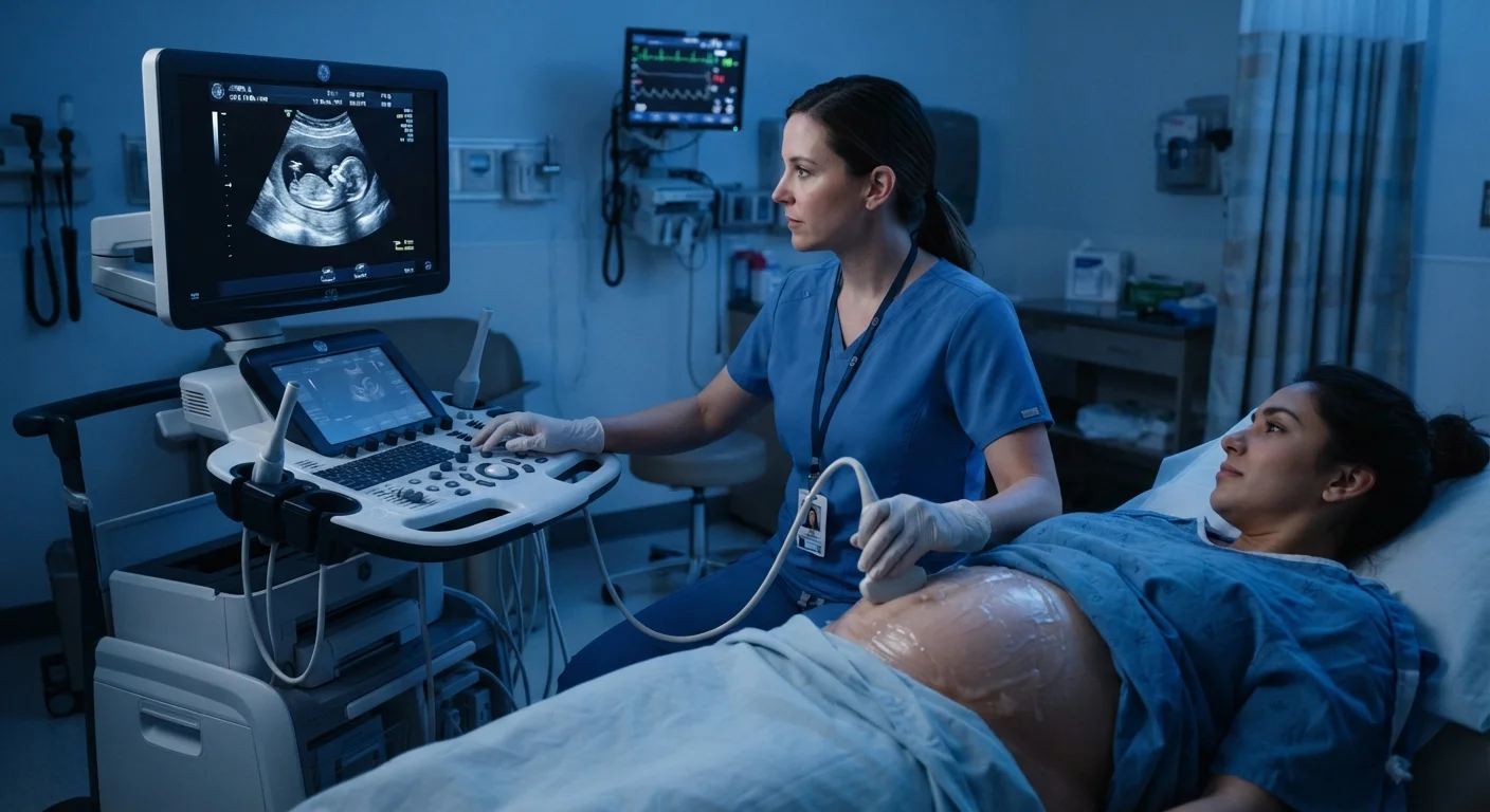
Photo: HerbHealWellness.com | Modern Guide To Wellness editorial
Key Takeaways
- Diagnostic ultrasound uses sound waves, not radiation, to visualize internal structures in real time.
- It is used across cardiology, emergency medicine, gastroenterology, vascular care, and musculoskeletal medicine — not just obstetrics.
- Different probe types and frequencies are selected depending on which organ or tissue is being examined.
- Ultrasound findings are interpreted alongside patient history and other test results, not in isolation.
- Point-of-care ultrasound allows bedside assessment, accelerating clinical decision-making in urgent settings.
How Ultrasound Creates Images
At its core, diagnostic ultrasound works on the same principle as sonar: sound waves are emitted, they interact with tissue, and the returning echoes are analyzed. The transducer — the handheld probe a clinician moves across the skin — both transmits and receives these sound pulses. Different tissues reflect sound differently: dense structures like bone produce strong echoes (appearing bright white on screen), while fluid-filled structures such as the bladder or a cyst transmit sound easily and appear dark.
The computer processes thousands of returning echoes per second to render a moving, real-time image called a sonogram or echogram. This live display is one of ultrasound's most practical advantages — clinicians can observe organ motion, valve function, and blood flow dynamics as they happen. Specialized modes extend this further: Doppler ultrasound detects the direction and velocity of blood flow by measuring shifts in sound frequency, which is essential in vascular and cardiac assessment.
For a broader look at how imaging technologies fit into clinical practice, see our plain-language tour of diagnostic lab equipment.
~40%
Proportion of global imaging studies using ultrasound
The World Health Organization has identified ultrasound as one of the most widely used medical imaging modalities worldwide, partly due to its portability and absence of ionizing radiation.
2–18 MHz
Typical frequency range of diagnostic ultrasound probes
Higher-frequency probes are used for superficial structures like tendons; lower frequencies are needed to image deeper organs such as the liver or kidneys.
>50 years
Duration of routine clinical ultrasound use
Real-time B-mode (brightness-mode) ultrasound became established in clinical medicine during the 1970s, building on earlier pulse-echo research.
Clinical Applications Beyond Obstetrics
Pregnancy imaging is perhaps the most publicly recognized use of ultrasound, but it represents only a fraction of its clinical scope. Across hospital departments, ultrasound informs decisions in conditions that affect virtually every major organ system.
- Cardiology (Echocardiography): A specialized cardiac ultrasound — the echocardiogram — evaluates heart chamber size, wall motion, valve function, and ejection fraction. It is indispensable in diagnosing heart failure, valve disease, and pericardial effusion (fluid around the heart).
- Abdominal imaging: Clinicians use ultrasound to identify gallstones, assess liver texture for fatty change or cirrhosis, measure kidney size, and detect abdominal aortic aneurysm.
- Vascular medicine: Duplex ultrasound combines conventional imaging with Doppler to map blood flow in arteries and veins, detect deep vein thrombosis (DVT), and assess carotid artery stenosis — a risk factor for stroke.
- Emergency medicine: Portable ultrasound enables rapid bedside evaluation for internal bleeding, pneumothorax (collapsed lung), and cardiac tamponade in trauma settings.
- Musculoskeletal care: Tendons, muscles, ligaments, and bursae are readily visualized with high-frequency probes, guiding both diagnosis and injection procedures for conditions like rotator cuff tears or plantar fasciitis.
- Thyroid and neck imaging: Ultrasound characterizes thyroid nodules and cervical lymph nodes, often directing subsequent biopsy decisions.
Ultrasound in Context: What It Can and Cannot Do
Ultrasound excels at imaging soft tissue and fluid, but it has well-recognized limitations. Sound waves do not penetrate bone well, making brain and spinal cord imaging impractical without specialized approaches. Gas within the bowel scatters sound, limiting visibility of structures behind it. Patient body habitus and the operator's technical skill also influence image quality — ultrasound is notably operator-dependent, meaning results can vary between practitioners.
Clinicians therefore integrate ultrasound findings with patient history, physical examination, laboratory results, and sometimes additional imaging modalities. No single scan provides a complete picture. As explained in our article on why imaging alone cannot diagnose most conditions, context is essential to reaching an accurate clinical conclusion.
It is also worth noting that diagnostic ultrasound is distinct from therapeutic ultrasound, which uses sound energy to heat deep tissue rather than to create images. These are separate technologies used for different purposes — a distinction explored in detail in our overview of how ultrasound therapy differs from diagnostic scans.
Ask Your Clinician Which Probe Is Being Used
This article is for general informational purposes only and does not constitute medical advice. Always consult a qualified healthcare professional regarding any symptoms, diagnoses, or treatment decisions.
Frequently Asked Questions
The content on this site is for informational purposes only and is not a substitute for professional advice. Always consult a qualified professional for guidance specific to your situation.