
Age-Related Macular Degeneration: A Complete Overview of AMD

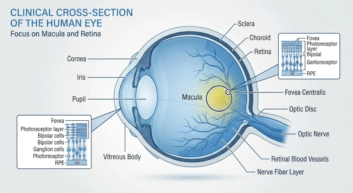
Photo: HerbHealWellness.com | Modern Guide To Wellness editorial
Key Takeaways
- AMD is the leading cause of irreversible central vision loss in adults over 50 in the United States.
- Dry AMD progresses gradually through drusen accumulation; wet AMD can cause rapid, severe vision loss.
- Early AMD often has no symptoms — routine eye exams are essential for detection.
- Age, smoking, and family history are among the strongest modifiable and non-modifiable risk factors.
- An Amsler grid is a simple at-home monitoring tool recommended for people diagnosed with AMD.
- There is no cure for AMD, but treatment options exist for wet AMD that can slow progression.
What Is Age-Related Macular Degeneration?
Age-related macular degeneration (AMD) is a progressive eye condition affecting the macula — the small, central portion of the retina responsible for sharp, detailed vision. The retina lines the back of the eye and converts light into signals sent to the brain. When the macula deteriorates, tasks that require fine detail — reading, recognizing faces, or driving — become increasingly difficult.
AMD is the leading cause of irreversible central vision loss among adults aged 50 and older in the United States. Importantly, AMD does not affect peripheral (side) vision, meaning complete blindness is not typical — but significant functional impairment is common in advanced stages.
There are two main forms of AMD: dry AMD (also called non-neovascular AMD), which accounts for roughly 80–90% of cases, and wet AMD (neovascular AMD), which is less common but responsible for the majority of severe vision loss associated with the condition. Understanding both forms is essential to recognizing how the disease can progress. For a broader look at how aging affects the eyes, see our guide on protecting eye health as you age.
11 million+
Americans affected by AMD
The American Academy of Ophthalmology estimates that more than 11 million Americans have some form of AMD.
2–4×
Increased AMD risk from smoking
Multiple large studies have found that current smokers face two to four times the AMD risk compared to non-smokers.
80–90%
AMD cases classified as dry form
The vast majority of AMD diagnoses are the dry (non-neovascular) type, though wet AMD causes most severe vision loss.
Dry AMD: Stages and What Drusen Deposits Mean
Dry AMD is characterized by the accumulation of drusen — small yellowish deposits of protein and lipid material — beneath the retina. Drusen are graded by size and number, and their characteristics help clinicians classify AMD into three stages:
- Early AMD: Small to medium drusen, no vision loss, and typically no symptoms. Often discovered incidentally during a routine dilated eye exam.
- Intermediate AMD: Larger drusen or pigment changes in the retinal pigment epithelium (RPE). Mild vision problems may begin, such as difficulty in low light.
- Late (Advanced) Dry AMD: Also called geographic atrophy — areas of the RPE and overlying photoreceptors break down, leading to noticeable central vision loss.
Because early and intermediate dry AMD typically produce no pain and limited visual symptoms, many people are unaware of its presence. This mirrors a broader pattern of silent eye disease progression described in our article on why eye conditions often have no early symptoms.
Check Your Vision Regularly at Home
Wet AMD: When New Blood Vessels Threaten Vision
Wet AMD occurs when abnormal new blood vessels grow beneath the retina — a process called choroidal neovascularization (CNV). These vessels are fragile and prone to leaking fluid or blood, which can rapidly distort or destroy macular tissue. Unlike dry AMD's gradual decline, wet AMD can cause significant central vision loss within days to weeks.
Symptoms that may signal wet AMD include:
- Straight lines appearing wavy or distorted (metamorphopsia)
- A dark, blurry, or blank spot in the center of vision
- Colors appearing less vivid
- Sudden or rapid change in visual clarity
Wet AMD Requires Prompt Medical Attention
It is worth noting that wet AMD almost always develops in eyes that already have dry AMD. Any person with a diagnosis of intermediate or advanced dry AMD should be counseled to monitor their vision closely and seek prompt care if changes occur.
Risk Factors You Should Know
AMD has several well-established risk factors, some modifiable and others not:
- Age
- The risk rises sharply after age 50 and increases significantly with each subsequent decade.
- Smoking
- Current smokers face approximately two to four times the risk of AMD compared to non-smokers — one of the strongest modifiable risk factors identified in research.
- Family history and genetics
- Variants in several genes, including CFH and ARMS2, are associated with significantly elevated AMD risk.
- Race and ethnicity
- AMD disproportionately affects non-Hispanic white adults, though it can occur in all populations.
- Cardiovascular risk factors
- Hypertension and high cholesterol have been associated with increased AMD risk in some studies.
- Light iris color and sun exposure
- People with lighter-colored eyes may have somewhat higher susceptibility, and cumulative UV exposure is a contributing factor.
Diet also plays a supporting role. Research has examined how specific nutrients — particularly antioxidants and omega-3 fatty acids — relate to AMD risk and progression. For a thorough review of what science says, see our article on the role of nutrition in eye health.
If you have intermediate AMD, ask your eye doctor whether the AREDS2 vitamin formulation is appropriate for you — it was specifically studied in people at elevated risk of progression to advanced AMD.
Never dismiss sudden visual distortion or a new blank spot as normal aging — these changes warrant a same-day or next-day call to your eye care provider.
How AMD Is Diagnosed and Monitored
AMD diagnosis typically involves several components performed by an ophthalmologist or optometrist:
- Dilated fundus examination: The eye is dilated so the clinician can inspect the retina and macula directly for drusen, pigment changes, or atrophy.
- Optical coherence tomography (OCT): A non-invasive imaging test that produces detailed cross-sectional images of the retina, allowing precise assessment of retinal layers and fluid accumulation.
- Fundus photography: Retinal photos document baseline appearance and allow comparison over time.
- Fluorescein angiography: A dye-based imaging test used to detect and map abnormal blood vessel growth in suspected wet AMD.
For people already diagnosed with AMD, Amsler grid testing is commonly recommended as an at-home monitoring tool. The grid is a simple pattern of straight lines with a central dot; distortion or missing areas in the grid can signal progression and should prompt a call to an eye care provider promptly.
Routine comprehensive eye exams remain the cornerstone of AMD detection — particularly in the early, symptom-free stages. The Eye Exam & Prevention hub offers further guidance on screening recommendations and preventive habits.
Living With AMD: What to Expect
An AMD diagnosis is understandably concerning, but the trajectory varies widely between individuals. Many people with early or intermediate dry AMD retain functional vision for years or decades. Those with advanced AMD can benefit from low vision rehabilitation — a specialty that helps individuals adapt to vision changes using magnification devices, improved lighting, and practical daily living strategies.
For wet AMD, clinicians commonly use anti-VEGF (vascular endothelial growth factor) injections delivered directly into the eye to slow or stabilize abnormal blood vessel growth. These treatments have substantially changed outcomes for wet AMD since their introduction. Photodynamic therapy is another approach used in certain cases. Treatment decisions are individualized and should always be discussed directly with a retinal specialist or ophthalmologist.
AMD is one of several serious conditions that may coexist in older adults. Understanding how it differs from other retinal conditions — such as diabetic retinopathy or conditions like cataracts — helps readers and clinicians make well-informed decisions about care priorities.
“The earlier we identify macular degeneration, the more options we have to help patients protect their remaining vision and adapt to any changes.”
— Paul Sieving, Former Director, National Eye Institute, National Institutes of Health
This article is for informational purposes only and does not constitute medical advice. If you have concerns about your vision or eye health, consult a qualified eye care professional.
The content on this site is for informational purposes only and is not a substitute for professional advice. Always consult a qualified professional for guidance specific to your situation.