
Eye Care for People with Diabetes: Why the Stakes Are Higher

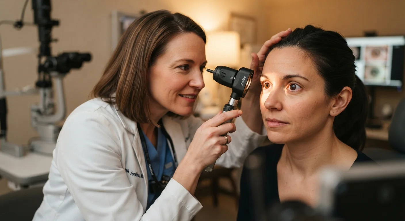
Photo: HerbHealWellness.com | Modern Guide To Wellness editorial
Key Takeaways
- Diabetic eye disease can cause irreversible vision loss before any symptoms appear.
- Annual dilated eye exams are recommended for most people living with diabetes.
- Early detection through routine screening significantly improves treatment outcomes.
- Blood sugar, blood pressure, and cholesterol control all influence eye health in diabetes.
- A dilated exam goes far beyond checking your prescription — it inspects the retina directly.
Why Diabetes Changes the Rules for Eye Care
For most adults, a routine eye exam every year or two is a reasonable baseline. For people living with diabetes, that calculus shifts considerably. Elevated blood sugar, over time, damages the tiny blood vessels that nourish the retina — the light-sensitive tissue at the back of the eye. This damage can begin silently, long before any change in vision is noticeable.
As our colleagues explain in their overview of why serious eye conditions often have no early symptoms, the absence of discomfort is not reassurance. Diabetic eye disease is one of the clearest examples: by the time blurry vision or floaters appear, meaningful retinal injury may already have occurred. This is why diabetes fundamentally changes the standard of care for vision health.
Diabetes Is a Systemic Condition
Conditions People with Diabetes Are at Higher Risk For
Diabetes raises the risk of several distinct eye conditions, not just one.
Retina
A thin layer of light-sensitive tissue at the back of the eye that converts light into signals sent to the brain. It is the structure most directly affected by diabetic eye disease.
Diabetic retinopathy
Damage to the blood vessels in the retina caused by prolonged elevated blood sugar. It can progress silently and is a leading cause of preventable vision loss in working-age adults.
Macular edema
Swelling in the macula — the central part of the retina — due to fluid leaking from damaged blood vessels. It causes blurring of central, detailed vision.
Dilated eye exam
An eye examination in which drops are used to widen the pupil, allowing the eye doctor to view the retina and optic nerve fully. It is the standard method for screening diabetic eye disease.
Intraocular pressure
The fluid pressure inside the eye. Elevated pressure is a key risk factor for glaucoma and is routinely measured during comprehensive eye exams.
Optical coherence tomography (OCT)
An imaging technology that creates detailed cross-sectional pictures of the retina's layers. It helps eye doctors detect and monitor subtle changes in retinal structure.
- Diabetic retinopathy is the most common and well-studied complication. Chronically elevated blood sugar weakens and distorts the retinal blood vessels, causing them to leak or grow abnormally. For a detailed breakdown of how it progresses stage by stage, see our article on how elevated blood sugar affects the eyes.
- Diabetic macular edema (DME) occurs when fluid accumulates in the macula — the central region responsible for sharp, detailed vision — reducing visual acuity.
- Cataracts tend to develop earlier and progress faster in people with diabetes than in the general population.
- Glaucoma risk is also modestly elevated, adding another reason why comprehensive exams matter.
Each of these conditions has distinct characteristics, but they share a common thread: earlier detection leads to more treatment options and better outcomes.
What a Diabetic Eye Exam Actually Involves
Many people are surprised to learn how much information a thorough eye exam can reveal. As detailed in our article on what your eye doctor can infer beyond your prescription, a dilated exam allows the clinician to directly examine the retina, optic nerve, and blood vessels — structures invisible without dilation.
During a diabetic eye exam, you can typically expect:
- Visual acuity testing — the standard letter chart to assess current sharpness of vision.
- Pupil dilation — eye drops widen the pupils, giving the examiner an unobstructed view of the retina. Vision may remain blurry and light-sensitive for several hours afterward, so plan transportation accordingly.
- Retinal examination — the doctor looks for early signs of vessel damage, leakage, abnormal new vessel growth, and swelling.
- Intraocular pressure measurement — screens for glaucoma risk.
- Imaging — many practices use retinal photography or optical coherence tomography (OCT) to capture detailed images of retinal layers for monitoring changes over time.
Plan Ahead for Your Dilation Appointment
How Often Should You Have Your Eyes Examined?
General guidance from major ophthalmologic organizations recommends that adults with diabetes have a comprehensive dilated eye exam at least once a year. For those newly diagnosed with type 2 diabetes, a baseline exam shortly after diagnosis is advised, since blood sugar may have been elevated for some time before diagnosis. People with type 1 diabetes are typically advised to begin annual exams within a few years of diagnosis.
Your individual schedule may differ based on findings at previous exams. If early retinal changes are detected, more frequent monitoring — every three to six months — may be recommended. Conversely, if multiple consecutive exams show no signs of disease, your eye care provider might adjust the interval. Always follow the schedule your clinician recommends rather than a general rule.
Do Not Skip Exams When Vision Seems Fine
Keeping Your Eyes Healthier Between Appointments
Eye exams are essential, but what happens between visits also matters. The factors that drive diabetic eye disease are largely systemic, meaning your overall metabolic health directly influences your eye health.
- Blood sugar management — sustained high glucose levels are the primary driver of retinal vessel damage. Work with your diabetes care team to optimize control.
- Blood pressure — hypertension accelerates retinal vessel damage, compounding the effects of high blood sugar.
- Cholesterol — elevated lipids are associated with more severe forms of diabetic macular edema.
- Not smoking — smoking worsens vascular disease throughout the body, including the retina.
If any sudden changes in vision occur — new floaters, flashes of light, a shadow across your visual field, or a rapid change in clarity — contact an eye care professional promptly rather than waiting for your next scheduled appointment.
For a broader perspective on how vision changes over a lifetime and what to watch for, our guide on protecting eye health as you age offers useful context.
This article is for general informational and educational purposes only and does not constitute medical advice. Always consult a qualified healthcare professional for guidance specific to your health situation.
Frequently Asked Questions
The content on this site is for informational purposes only and is not a substitute for professional advice. Always consult a qualified professional for guidance specific to your situation.