
Everything Your Eye Doctor Can Infer Beyond Your Prescription

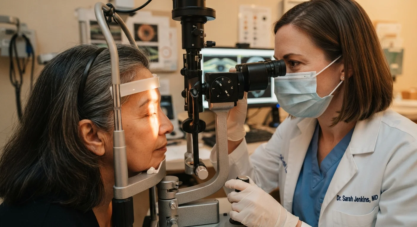
Photo: HerbHealWellness.com | Modern Guide To Wellness editorial
Key Takeaways
- Eye exams can detect early signs of diabetes, high blood pressure, and certain neurological conditions.
- The retina is the only place in the body where blood vessels can be observed directly without surgery.
- Dilated eye exams provide the most complete view of the optic nerve, retina, and internal eye structures.
- Many sight-threatening and systemic conditions show no symptoms in early stages, making routine exams essential.
- Share your full medical history and medications with your eye doctor — it shapes how they interpret findings.
The Eye as a Window to Whole-Body Health
Most people schedule an eye exam when they notice blurred vision or need a new prescription. But for optometrists and ophthalmologists, examining your eyes is also a systematic scan for broader health conditions — many of which have no obvious symptoms until significant damage has occurred.
The retina is uniquely revealing: it is the only location in the human body where blood vessels, nerve tissue, and circulation can be observed directly and non-invasively. Changes in those structures can reflect events happening elsewhere — in the cardiovascular system, the brain, or the metabolic system. Understanding what your eye doctor is looking for reframes a routine visit as something much more consequential.
For a deeper look at how ageing specifically changes what clinicians look for, see our guide on protecting eye health as you age.
What Happens During a Comprehensive Eye Exam
A comprehensive eye exam typically involves several distinct assessments beyond reading an eye chart. Your clinician will evaluate visual acuity, eye alignment, intraocular pressure (the fluid pressure inside the eye), and the health of the anterior segment — the front portion of the eye including the cornea, lens, and iris.
The most diagnostically rich portion is the dilated fundus examination. Dilating drops temporarily widen your pupils, allowing the clinician to examine the optic nerve head, the macula, the peripheral retina, and the retinal blood vessels in full detail. This step is not optional for a truly comprehensive assessment.
Always Ask About Dilation
Depending on your age, risk factors, and history, your doctor may also use additional tools such as optical coherence tomography (OCT) — a non-invasive imaging technology that produces cross-sectional maps of retinal layers — or visual field testing, which assesses peripheral vision and can detect early glaucoma or neurological changes.
If you're unsure whether your current provider performs comprehensive exams, our article on optometrist vs. ophthalmologist differences can help clarify scope of practice.
Systemic Conditions Detected Through the Eyes
90%
Of diabetic retinopathy vision loss is preventable
According to the American Academy of Ophthalmology, timely detection and treatment can prevent the majority of severe vision loss from diabetic retinopathy.
3M+
Americans living with glaucoma, many undiagnosed
The Glaucoma Research Foundation estimates that over half of people with glaucoma in the US are unaware they have the condition.
1 in 3
Adults with vision loss have not had an eye exam in a year
CDC data suggests a substantial proportion of American adults with vision impairment have not had a recent comprehensive eye examination.
Several serious systemic conditions leave early, identifiable marks on ocular tissue — often before a patient experiences any general symptoms.
Diabetes and Diabetic Retinopathy
Elevated blood sugar damages small blood vessels throughout the body, and the retina's fine capillaries are among the first affected. Clinicians look for microaneurysms (tiny bulges in vessel walls), retinal hemorrhages, and abnormal new vessel growth — hallmarks of diabetic retinopathy. In some cases, eye exams have led to a first-ever diabetes diagnosis. For those already managing diabetes, eye care for people with diabetes warrants particular attention, and understanding diabetic retinopathy's stages is important context.
Hypertension
Chronic high blood pressure causes distinctive changes to retinal arteries — they may appear narrowed, take on a copper or silver wire appearance, or show signs of arteriovenous nicking (where arteries compress adjacent veins at crossing points). These findings, collectively termed hypertensive retinopathy, can prompt a clinician to recommend blood pressure evaluation when a patient has no prior diagnosis.
High Cholesterol
Deposits of cholesterol-rich plaques called Hollenhorst plaques can become lodged in retinal arteries and are visible on fundoscopic examination. Their presence may indicate embolic disease in the carotid arteries, prompting further cardiovascular workup.
Eye Findings Are a Starting Point, Not a Diagnosis
Neurological and Vascular Warning Signs
The optic nerve connects the eye directly to the brain, which makes it a critical structure for detecting neurological disease.
Glaucoma
Glaucoma causes characteristic changes to the optic nerve head — particularly a condition called cupping, where the central depression of the nerve enlarges as nerve fibers are lost. Because peripheral vision is affected first, patients rarely notice symptoms until the disease is advanced. Intraocular pressure measurement and optic nerve imaging are standard elements of detection. Browse common eye conditions for more on glaucoma and related diagnoses.
Intracranial Pressure and Tumors
Swelling of the optic disc — called papilledema — can indicate dangerously elevated pressure inside the skull, caused by conditions ranging from idiopathic intracranial hypertension to brain tumors. An eye doctor who identifies papilledema will refer immediately for urgent neurological evaluation.
Multiple Sclerosis
Inflammation of the optic nerve (optic neuritis) is frequently an early presentation of multiple sclerosis. Reduced color perception, pain with eye movement, or sudden vision changes in one eye can prompt referral to neurology.
If you notice even brief or transient changes in vision — a few seconds of darkness in one eye, sudden floaters, or a loss of peripheral vision — report them at your exam even if they resolved. These episodes can signal vascular events that warrant urgent evaluation.
Bring a list of all medications you take, including vitamins and herbal supplements — some have documented effects on the retina, optic nerve, or intraocular pressure that your eye doctor needs to account for.
Additionally, changes in how the pupils respond to light — called pupillary light reflex abnormalities — can signal damage anywhere along the visual pathway, from the eye to the brain.
How to Get the Most From Your Next Exam
The diagnostic value of an eye exam depends significantly on the information you bring to it. Your clinician needs to know your full medical history, current medications (including supplements), family history of eye disease, and any symptoms — even transient ones like brief double vision or intermittent flashes of light.
Our preparation checklist, before your eye appointment, covers exactly what to bring and discuss. The more context your provider has, the more precisely they can interpret what they see.
Don't Skip Exams Without Symptoms
Frequency matters too. For most healthy adults, a comprehensive dilated exam every one to two years is a reasonable baseline — but your provider may recommend more frequent visits if you have diabetes, a family history of glaucoma, or other risk factors. Children, older adults, and those with chronic health conditions should discuss individualized schedules with their care team.
If you experience symptoms like sudden vision loss, persistent floaters, light flashes, or significant changes in vision, do not wait for your scheduled visit — seek care promptly. And if you're managing screen-related discomfort, our article on digital eye strain separates established fact from common misconceptions.
This article is for informational purposes only and does not constitute medical advice. Always consult a qualified eye care professional or physician regarding any personal health concerns, symptoms, or decisions about your care.
The content on this site is for informational purposes only and is not a substitute for professional advice. Always consult a qualified professional for guidance specific to your situation.