
What Actually Happens During a Routine Eye Exam

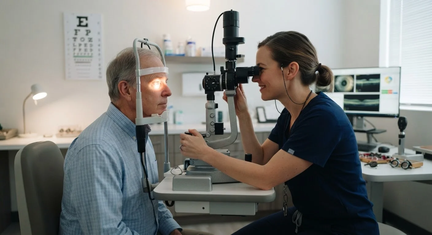
Photo: HerbHealWellness.com | Modern Guide To Wellness editorial
Key Takeaways
- A routine eye exam checks both your vision clarity and the structural health of your eyes.
- Eye pressure testing screens for glaucoma, which often has no early symptoms.
- Dilation allows your doctor to inspect the retina and optic nerve directly.
- Many systemic conditions, including diabetes and hypertension, can show early signs in the eye.
- Most adults should have a comprehensive eye exam at least every two years, or more frequently based on risk.
The Exam Starts Before the Instruments Come Out
When you arrive, a staff member or technician will review your medical and ocular history before the doctor enters the room. This intake step — often underestimated — is where your doctor learns about current medications, family history of eye disease, and any symptoms you've noticed, such as blurred vision, floaters, or headaches.
Being thorough here pays dividends. Conditions like glaucoma, macular degeneration, and diabetic retinopathy all carry hereditary or systemic risk factors that shape how your exam is conducted. See our preparation checklist for what to bring and what details matter most.
Make the Most of Your History Review
Visual Acuity and Refraction: Finding Your Prescription
The familiar letter chart — formally called a Snellen chart — measures visual acuity, or how clearly you see at a standard distance. You'll read progressively smaller rows with each eye separately. This establishes your baseline and flags any reduction since your last visit.
Next comes refraction, the process that determines your lens prescription. Your doctor uses a device called a phoropter — the large instrument you look through while comparing lens options — to identify the combination of corrective lenses that gives you the sharpest vision. Automated pre-testing with a refractometer often precedes this step to give the doctor a starting point.
It's worth knowing that a vision screening at school or work only approximates this step and cannot substitute for it. The difference between a screening and a full exam is significant in terms of what can be detected and how precisely.
Eye Pressure, Alignment, and the Slit Lamp
Intraocular pressure (IOP) measurement — tonometry — screens for elevated pressure associated with glaucoma. The air-puff method is common in primary care settings; contact tonometry, using a gentle probe after numbing drops, is considered more precise. Elevated pressure alone doesn't confirm glaucoma, but it flags patients for closer monitoring.
Your doctor will also assess eye muscle coordination and alignment using a penlight or cover test, checking that both eyes move together and focus accurately. This detects strabismus (eye misalignment) and binocular vision problems that can cause headaches or reading difficulty.
The slit lamp is a microscope that illuminates and magnifies the front structures of the eye — eyelids, cornea, iris, and lens — under high magnification. This is how early cataracts, corneal irregularities, and signs of dry eye disease are identified.
Not All Exams Include Every Test
Dilation and the View Inside Your Eye
Dilating drops temporarily widen your pupils, giving your doctor an unobstructed view of the retina, macula, and optic nerve — structures at the back of the eye that cannot be fully examined otherwise. The doctor uses an ophthalmoscope or a condensing lens with the slit lamp to scan these tissues.
This is arguably the most clinically significant part of the exam. Changes to retinal blood vessels may suggest diabetes or hypertension. A pale or cupped optic nerve can indicate glaucoma. Drusen deposits in the macula are associated with age-related macular degeneration. For a deeper look at what these findings mean, see everything your eye doctor can infer beyond your prescription.
93 million
U.S. adults at high risk for vision loss
According to the CDC, about 93 million U.S. adults are at high risk for serious vision loss, but only half visited an eye doctor in the past 12 months.
50%
Glaucoma cases estimated to be undiagnosed
The Glaucoma Research Foundation estimates that approximately half of people with glaucoma in the U.S. are unaware they have the condition, often due to its lack of early symptoms.
Dilation causes temporary light sensitivity and blurred near vision lasting two to six hours. Bringing sunglasses is practical; arranging a driver is worth considering.
After the Exam: Understanding Your Results
Your doctor will discuss findings at the end of the visit — your prescription, any observed changes in eye health, and whether follow-up or referral is needed. If your eyes are healthy and stable, you'll leave with a prescription and a recommended return interval.
How often that interval should be depends on your age and individual risk profile. The recommended exam frequency by age and risk factor is a useful reference for planning ahead. Early detection remains the most effective strategy for conditions like glaucoma and macular degeneration, which can progress silently before symptoms appear. Routine exams, even when vision feels fine, are a core part of preserving long-term eye health.
This article is for general informational and educational purposes only and does not constitute medical advice. Consult a qualified eye care professional for guidance specific to your health and vision needs.
Frequently Asked Questions
The content on this site is for informational purposes only and is not a substitute for professional advice. Always consult a qualified professional for guidance specific to your situation.